
George Mason team earns $2.1M to study seizure dynamics
The National Institutes of Health (NIH) awarded a George Mason University research team $2.1 million to create a system that could revolutionize the study of seizures.
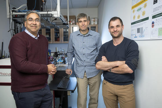
The project is led by Principal Investigator (PI) Rob Cressman, Associate Professor in the Department of Physics and Astronomy, with co‑Principal Investigators Parag Chitnis and Remi Veneziano from the Department of Bioengineering. Together, the research team will develop an integrated imaging and sensing system designed to identify regions of the brain exhibiting dysregulation of potassium ion activity.
“Potassium is one of those ions that actually inhibits activation of cells. If there's a dysfunction in those ions, you get runaway signaling. A seizure is an example of that,” said Cressman, who has studied epilepsy since his postdoctoral work. “We’re trying to develop tools that help us understand that better.”
When the biocompatible nanosensors, made of DNA and light-sensitive dye, encounter a local change in potassium concentration, they change their shape resulting in a change in their near-infrared fluorescence signal.
“We want to detect even very small changes in potassium,” said Veneziano, whose work involves synthesizing new nanomaterials from DNA particles, proteins, and lipids. “The system we’ve built is modular, versatile, and highly sensitive.”
These signals can be mapped in real-time using a near-infrared imaging device to understand dynamic changes in ion concentration.
One of the most promising applications is image-guided surgical intervention for epilepsy. By pinpointing seizure foci through potassium mapping, surgeons may be able to perform precise tissue ablation, removing only the affected areas.
“If you can identify the focus and remove that part of the tissue, you can sometimes stop the seizures,” said Cressman.
The four-year NIH-funded project, previously funded by the National Science Foundation (NSF), builds on years of research supported by George Mason’s Center for MedTech Innovation.
“This grant allows us to translate our NSF-funded feasibility work into real-world applications,” said Chitnis, who studies brain-body interactions using light and ultrasound. “We’re not just studying disease—we’re building tools that could guide future interventions.”
The toolkit will be tested across multiple platforms—from cell cultures to brain slices to intact mammalian models—allowing researchers to study bioelectrical activity at various scales.
The team also sees potential for assessing functional recovery after traumatic brain injury. Scar tissue can disrupt potassium regulation, and the nanosensors may help identify regions at risk of developing seizure foci. As the research progresses, the team hopes to open new pathways for understanding and treating complex brain disorders.